“I will not go out for a CT scan”, said my Mom whom doctor referred CT scan so that the reason for her sharp pains in the body is discovered. Being terrified of the doughnut-shaped machine to go in, she was talking to my cousin who is a doctor. Initially he gave some comic remarks for such kind of fear but after a few minutes, the conversation turned its way to a serious one. And he told that we can get a 3D model from 2D X-ray images of a bone from a patient’s body.
However, Mom’s fear was gone away to some extent because the way he made us know was alternative to CT scan. Here is the essence of that conversation that will be quite informative for you.
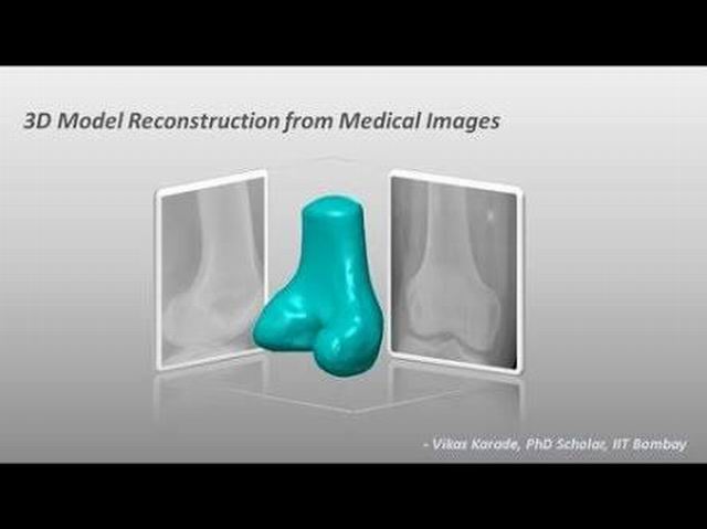
A software from OrthoCAD lab can make a 3D model from 2D X-ray using 2D X-ray images. The process takes only a few minutes.
Yes, you are not wrong if thinking of the 2D views that can be generated from a 3D model. But to think that a 3D model can’t be generated by using 2D X-ray, you are wrong.
Dr. Manish Agarwal’s argument that if we can reconstruct the model in our mind, then it can also be programmed in the computer to get a 3D model from a 2D X-ray.
Reconstruction of the 3D model is also possible from a CT scan but in this process, several hundred times more radiations are required. So the process becomes more expensive for a patient.
A research scholar Vikas Karade and a Professor B. Ravi at OrthoCAD Lab followed the argument of Dr. Manish Agarwal and started their journey to reach the 3D model through a 2D X-ray.
The program that can be aptly named ‘XrayTo3D’ can convert 2D X-ray images of a bone from a patient into a 3D model.
To see the demo version of this program you can visit OrthoCAD lab website. Any clinician can use it freely.
Only once created a 3D template of corresponding bone and one or two X – ray images taken at perpendicular planes are required as input. The 3D template is created from CT images of a healthy patient and is projected on the two planes of the X – ray images.
When the projections are matched in shape with the X – ray images by the algorithm, it takes less than a minute to convert these projections back into 3D model.
This 3D model is created with an error less than 1.5 mm compared to that we reconstruct from CT images.
Orthopedic surgeons can use this program to plan surgeries in 3D without using radiation-intensive. It saves the patients from getting into the doughnut-shaped expensive CT scanners.
The rural areas where CT scan machines are rare can avail this software.
Would not it luck if implemented on the tablets and smartphones? Well, Vikas (Gandhian Young Technological Innovation Award holder of 2014) is working on this idea.


